
A view far wider than a standard retina photo
Your retina, the thin layer at the back of your eye, is where many eye diseases show up first. A standard retina photo shows only the center of it. Optos, the company that makes Optomap, says each Optomap image captures about 82% of the retina, around 200 degrees, compared with roughly 15% for a standard photo.
- Shows the center and the far edges of the retina in one image
- Reviewed with you on screen by Dr. Hidalgo
- Kept on file to compare year after year
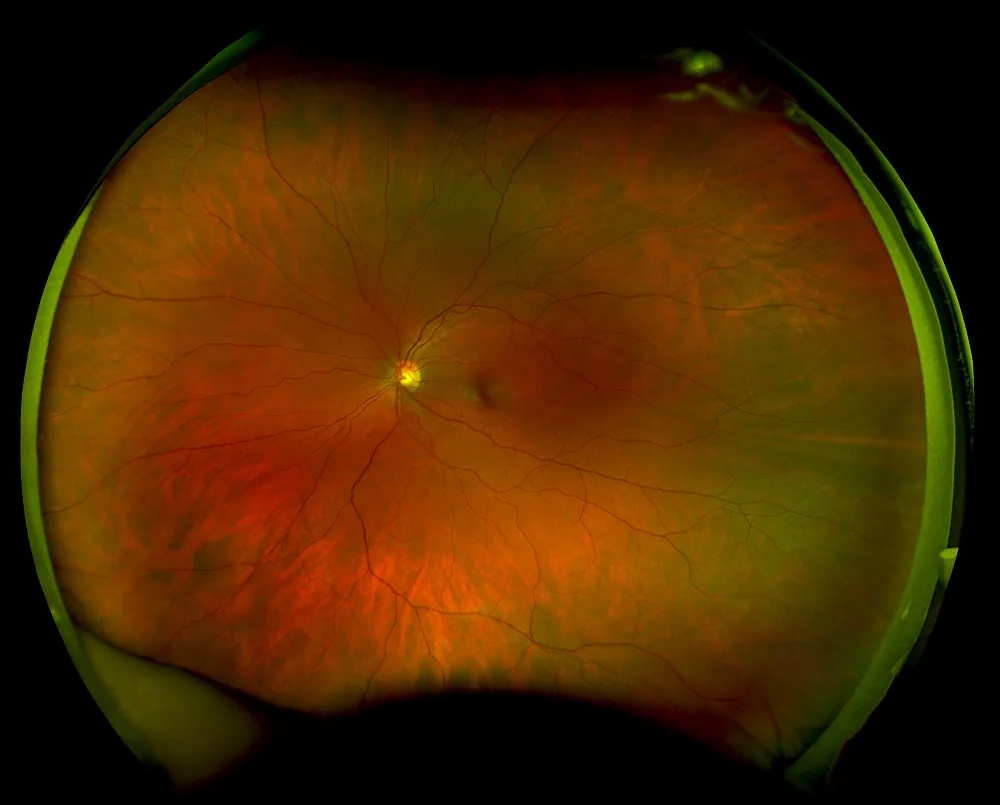
Often an alternative to dilating drops
Dilating drops widen your pupils so the doctor can see inside your eye, but they can leave your vision blurry and sensitive to light for hours. Because Optomap gives Dr. Hidalgo such a wide view of your retina, many patients can skip the drops and get right back to their day.
- No blurry, light-sensitive hours after your visit
- Drive yourself home and go back to work or school
- A wide view of your retina in under a second
Please note: whether you still need dilating drops is up to Dr. Hidalgo, based on your exam results. Some patients and eye conditions still call for a dilated exam, even with Optomap.
What Optomap helps Dr. Hidalgo look for
Many of these conditions cause no symptoms at first. Seeing them early gives you more options.
Diabetic eye disease
Changes in the tiny blood vessels of the retina, which can appear before your vision changes.
Glaucoma
Signs of damage to the optic nerve, which connects your eye to your brain.
Macular degeneration
Changes in the macula, the center of the retina you use to read and see faces.
Retinal tears and detachments
Problems near the outer edges of the retina, an area a standard view can miss.
The retina can also show signs of other health conditions, such as high blood pressure. Optomap is part of your eye exam, not a replacement for it.
Quick, easy and painless
- Look Into the Device
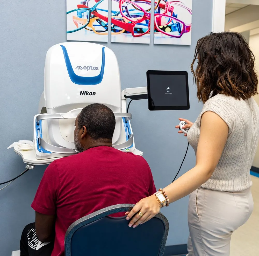
You sit at the Optomap and look into it, one eye at a time.
- One Quick Flash
Each image takes under a second. It's painless, and nothing touches your eye.
- Review It Together
Dr. Hidalgo goes over your images with you on screen and keeps them on file for next time.
Add it to your exam for $35
Insurance doesn't cover Optomap at our office, so it's always $35 out of pocket, whatever plan you have.
Common questions
Does Optomap hurt?
No. It's a quick flash of light, and nothing touches your eye.
Will I still need dilating drops?
Maybe not. Optomap is often an alternative to dilation, but Dr. Hidalgo decides based on your exam results, and some patients still need a dilated exam.
Does insurance cover Optomap?
No. At our office, Optomap is always a $35 out-of-pocket add-on, with or without insurance.
How often should I get Optomap?
Many patients add it to every yearly exam, so Dr. Hidalgo can compare your images over time and catch changes early.
Can I see my images?
Yes. Dr. Hidalgo reviews them with you on screen during your visit.